7/30/2021

Physiology of Urine Formation.


Introduction.

  • Urine is a waste product formed by the kidneys after filtration of the blood.

Composition of Urine:

  • Color: Yellow / Pale yellow.

  • pH: 6 to 7.5

  • Volume: 1 to 2 Liters / Day.

  • Odor: Aromatic, Strong ammoniacal on standing or concentrating.

  • Composition:

Normal Ingredients:

  • Water, Urea, Uric Acid, Creatinine, Ammonia, Sodium, Potassium, Chlorides, Sulphates, Phosphates etc.

Abnormal Ingredients:

  • Glucose: Diabetes mellitus.

  • Proteins: Kidney infection.    

  • Blood Components: Kidney Infection.

  • Ketone bodies: Ketosis.

  • Bile Pigments: Liver disorder, heavy RBC destruction.

  • Urine production is lowered during sleep and exercise

Physiology of Urine Formation

  • The main function of the kidney is to clean the plasma, the waste product formed in the process is called “Urine”.

  • The urine formed in kidneys is transported via ureters to the urinary bladder, the temporary urine storage site, urine is then excreted from the body by a process called “Micturition”.

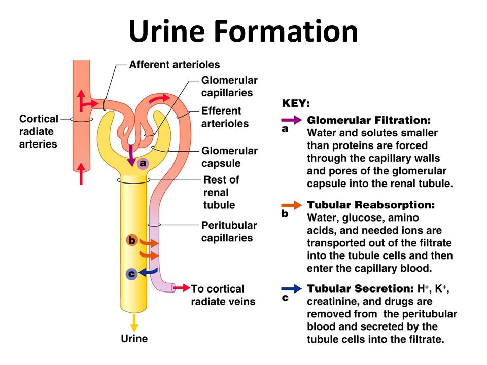
  • Urine is formed in the kidney in the following three steps,

  1. Glomerular Filtration

  2. Selective Reabsorption.

  3. Tubular Secretion

  1. Glomerular Filtration:

  • First step in the process of urine formation.

  • It occurs in the ‘renal corpuscle’ at the ‘filtration membrane’.

  • Afferent arteriole has a large diameter and efferent arteriole has a smaller diameter; this difference between diameters causes an increase in pressure inside glomerulus which facilitates filtration.

  • The blood cells, plasma proteins and any other larger molecules normally can not be filtered due to their larger size

  • The fluid that passes into the capsular space of the bowman's capsule is calledFiltrate”.

  • Glomerular Filtration Rate:

    • The amount of filtrate formed in all renal corpuscles of both the kidneys per minute is called “Glomerular Filtration Rate (GFR)”.

    • Normal GFR = 125 ml/min (125x60x24. 180 lit. per day)

  1. Selective Reabsorption:

  • The filtrate after entering the “renal tubule” gets reabsorbed.

  • About 99% of the filtrate is reabsorbed and enters the blood, while only 1% filtrate forms the urine.

  • The movement of water and other solutes from filtrate to peritubular capillaries is called “Selective Reabsorption.”

  •  The term “Selective” is used as only selected substances are reabsorbed e.g. glucose, amino acids, ions like sodium, chloride, potassium, bicarbonate, phosphate etc.

  • The reabsorption is active as well as passive, involving energy as well as without involving energy.

  • Selective reabsorption is influenced by various hormones like,

    • Parathyroid hormones (Parathyroid Gland): ↑ reabsorption of calcium and phosphate ions.

    • AntiDiuretic Hormone, ADH (Pituitary Gland): ↑ reabsorption of water.

    • Aldosterone (Adrenal Gland): ↑ Sodium and water reabsorption.

    • Atrial Natriuretic Peptide (Cardiac Atria): ↓ reabsorption of sodium and water from PCT.

  1. Tubular Secretion:

  • It is the third and last step in urine formation.

  • In this process various substances are added in filtrate, e.g. Potassium ions, Hydrogen ions, urea, ammonium ions, creatinine, certain drugs like Penicillin etc.

  • Tubular secretion of “Hydrogen Ions” is an important thing in relation to body pH maintenance.

Commonly asked Questions.

  1. What is Urine? Discuss physiology of formation of urine.

  2. With a well labelled diagram of Nephron discuss physiology of formation of urine.

  3. Draw a well labelled diagram of L. S. of kidney and discuss in detail the physiology of formation of urine.

Labels: ,

Nephron: Structural & Functional unit of Kidney.


Introduction.

  • Structural and functional unit of kidney.

  • Each kidney contains around “ One Millionnephrons.

  • Present in the renal cortex as well as in renal medulla

  • Two types:

    • Cortical Nephrons: 

      • Superficial.

      • Originate in the cortex and extend for a short distance in medulla.

      • Constitute around 80-90% of the total nephrons.

    • Juxtamedullary Nephrons:

      • Lie deeper.

      • Originate at the junction of medulla and cortex, go deeper in medulla.

      • Constitute around 10-20% of the total nephrons.

  • Each nephron consists of two units:

    • Renal corpuscle. (Filtration Unit) It is made up of,

      • Glomerulus.

      • Bowman's Capsule

    • Renal Tubule. (Transportation Unit) it is made up of,

      • Proximal Convoluted Tubule (PCT).

      • Loop of Henle.

      • Distal Convoluted Tubule. (DCT)

Renal Corpuscle:

  • It is a filtering chamber where plasma is filtered.

  • It has two components,

    • Glomerulus.

    • Bowman’s Capsule.

  • Glomerulus is a bunch of capillaries, blood enters in glomerulus via afferent arteriole and exits through efferent arteriole.

  • Afferent arteriole has a large diameter and efferent arteriole has a smaller diameter, this difference between diameters causes increase in pressure inside glomerulus which facilitates filtration.

  • Bowman's capsule is a funnel shaped part of nephron surrounding glomerulus.

  • Bowman's capsule’s outer wall called the parietal layer is separated from the inner wall called the visceral wall by a space called “Capsular Space”.

  • Bowman's capsule and glomerulus together form a “malpighian body”.

  • Juxtaglomerular apparatus is a specialized structure of cells that plays an important role in blood pressure maintenance

  • Filtration Membrane” is the contact point between glomerular capillaries and Bowman’s Capsule.

  • Blood from afferent arteriole enters glomerulus and is filtered i.e. blood cells, protein molecules are retained in capillary while plasma with solutes with smaller size escape from small pores on capillaries to capsular space and then to the renal tubule.

Renal Tubule:

  • It starts from a bowman's capsule and ends at collecting tubules.

  • Anatomically it is divided into three parts,

  1. Proximal Convoluted Tubule (PCT).

  2. Loop of Henle.

  • Ascending Loop of Henle.

  • Descending Loop of Henle.

  1. Distal Convoluted Tubule (DCT).

  1. Proximal Convoluted Tubule (PCT): 

  • It starts from Bowman’s capsule and continues down the descending loop of Henle.

  • It has an inner lining of cuboidal epithelium having a microvilli brush border (just like epithelial cells in the small intestine), which increases surface area for fluid absorption.

  • It absorbs ions like Na &  Cl, water and glucose.

  • It absorbs actively hence contain very large number of mitochondrias to produce ATPs

  1. Loop of Henle:

  • It is divided into two parts,

    • Descending Loop of Henle. (part going downwards)

    • Ascending Loop of Henle. (part going upwards)

  • It is present between PCT and DCT and has a sharp hairpin like structure.

  • It deals with maximum fluid absorption, it is more long in animals present in the desert.

  1. Distal Convoluted Tubule (DCT):

  • The highly coiled part starts after the ascending Loop of Henle.

  • It contains simple cuboidal epithelium.

  • It is shorter as compared to PCT.

  1. Collecting Duct:

  • DCT continues to form collecting ducts.

  • Collecting ducts are connected to many nephrons.

  • It contains columnar epithelium.

Commonly Asked Questions.

  1. Draw a well labeled diagram of the Nephron.

  2. With a well labelled diagram discuss the structure of Nephron.

  3. With a well labelled diagram of Nephron discuss the process of Urine formation.

Labels: