Tissue level of organization: Muscular Tissue.
Introduction:
Groups of cells having similar structure and performing similar functions are called “Tissue.”
Muscular tissue is specialized for contraction and brings movement.
There are three specialized muscular tissues present in the body as follows,
Skeletal Muscles.
Smooth Muscles.
Cardiac Muscles.
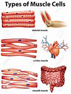
Skeletal Muscle:
The name is derived as they are attached to the skeleton and are responsible for skeletons movement or say Locomotion.
Also called “Striated muscle” or “Voluntary muscle”.
As they work under our will power they are called “Voluntary muscle”.
On Microscopic examination they appear striated due to precise arrangement of contractile proteins inside the cell.
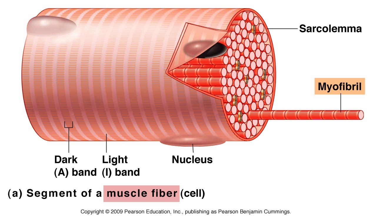
The plasma membrane and endoplasmic reticulum are special and hence called “Sarcolemma and Sarcoplasmic reticulum” respectively.
Muscle cells are also called “Myocytes” , “Muscle Fibers”.
Each muscle fiber contains many rod-like structures :Myofibrils.
Skeletal muscle cells are cylindrical in structure and contain many nuclei.
In the cell there is a typical arrangement of proteins responsible for contraction which gives striated appearance.
The impulses for muscle contraction generate at the brain or spinal cord and end at neuromuscular junctions.
Functions:
Movement.
Body posture.
Regulation of body temperature: Generate heat.
Smooth Muscle:
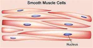
Also called “Non Striated Muscles” as they don't show striations.
Do not work under will power hence called as “Involuntary Muscles”.
They are present in internal organs hence called “Visceral Muscles.”
The cells contain a single central nucleus and are spindle shaped.
Contractions are slower and more sustained than skeletal muscles.
Functions:
Wall of blood vessels: Regulation of diameter.
Eye: Regulation of pupil size.
Intestine: Peristalsis.
Uterus: Contraction.
Cardiac Muscle:
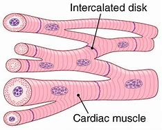
This tissue is only found in myocardium of the heart and hence called “Cardiac Muscle.”
This tissue when observed under a microscope shows striations like voluntary muscles but works like involuntary muscle hence it is considered as a special type of muscle.
The cells are branched and have a single nucleus.
The cell membrane at end is folded and fits in matching folds of adjacent cell membranes forming “intercalated discs”.
Intercalated disc is an important feature of cardiac muscle as it makes passage of electric impulses faster.
Heart beats on its own, the nerve supply only increases or decreases rate and force of contraction as per need of the situation.
Functions:
Contractions of heart.
Commonly Asked Questions.
Write a note on “Muscular Tissue.”
Draw a well labelled diagram of,
Cardiac Muscle.
Smooth Muscle.
Skeletal Muscle.