Cellular level of organization: Cell Division: Meiosis.
Introduction:
Cell is defined as a basic structural and functional unit of the body.
Cytology is a branch of science that deals with the study of cells.
The animal cell is divided into two major parts as,
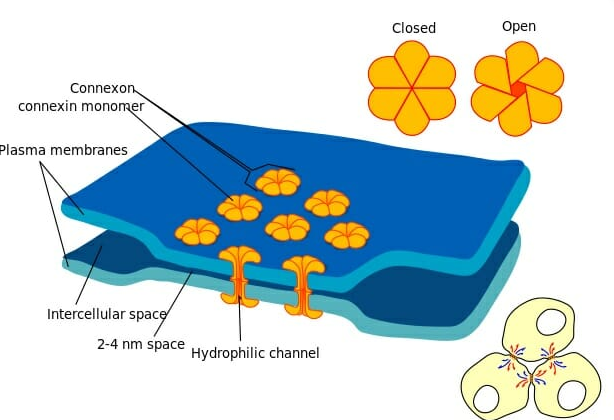
Plasma Membrane.
Subcellular organelles.
Nucleus.
Nucleolus
Mitochondria.
Golgi complex.
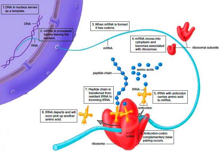
Ribosomes.
Endoplasmic reticulum.
Lysosomes.
Centrioles.
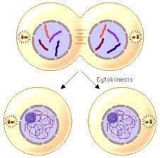
The cell cycle, or cell-division cycle, is the series of events that take place in a cell that cause it to divide into two daughter cells.
These events include the duplication of its DNA (DNA replication) and some of its organelles, and subsequently the partitioning of its cytoplasm and other components into two daughter cells in a process called cell division.

The events in the cell that include the duplication of its DNA (DNA replication) and some of its organelles, and subsequently the partitioning of its cytoplasm and other components into two daughter cells in a process called cell division.
Cell Division: The ability of the cell to produce daughter cells.
Normally, cells divide to replace dead or missing cells,
intestinal cells divide every 3 days, are broken down by digestion
blood cells last 3 months, are replaced by new cell division
nerve cells usually don't divide, last for life
Embryo: almost constant cell division. Every 30 minutes.
Sex cells are unique: in humans, males produce 500,000,000 /day. Females 1/month.
Meiosis.
Definition: It's a special type of cell division where the cell genome is reduced to half to produce “Four Haploid Cells”.
The cells produced in meiosis are called gametes.
As genetic material is reduced to half, it is also called as “Reductional Cell Division”.
Parent Cell (2n)-------> Daughter Cell (1n).
Meiosis I | Prophase I | Meiosis II |
Prophase I
| Leptotene
| Prophase II
|
Metaphase I
| 2) Zygotene | 2) Metaphase II |
Anaphase I
| 3) Pachytene | 3) Anaphase II |
Telophase I
| 4) Diplotene | 4) Telophase II |
Cytokinesis I
| 5) Diakinesis | 5) Cytokinesis II |
Meiosis I:
Prophase I:
Leptotene
Zygotene
Pachytene
Diplotene
Diakinesis.
Leptotene:
Also called “Leptonema” (Lepto= Thin, Nema= Thread).
In this stage condensation of Chromosomes takes place.
Chromosomes get converted into thin thread like structures.
Each maternal and paternal chromosome starts searching for their “homologous chromosomes”.

Zygotene:
Also called “Zygonema” (Zygo= Pair, Nema= Thread).
Maternal and Paternal Chromosomes come together to form a pair called “Synapse / Bivalent / Homologous pair”.

Pachytene:
Also called “Pachynema” (Pachy= Thick, Nema= Thread).
Most important event takes place i.e. “Crossing Over”.
Two nonsister chromatids overlap each other, the point of attachment called “Chiasmata”.
Due to this transfer of genetic material took place between maternal and paternal chromosomes.
Pachytene hence is an important step as the basis of evolution takes place here.

Diplotene:
Also called “Diplonema” (Diplo= Two, Nema= Thread).
Nuclear membranes and nucleolus start disappearing.
Centrioles get connected by spindle fibers.
Centrioles start moving to the opposite ends.
The overlapped chromosomes start repulsing each other.

Diakinesis:
Also called “Terminalization”.
Stage is characterized by the ending of cross over.
The repulsion between crossed over chromosomes continues, still there is attachment at tip.
Nuclear membranes and nucleolus disappear.
Centrioles got connected by spindle fibers.
Centrioles move to opposite ends.
This stage marks the end of Prophase I.

2) Metaphase I:
Chromosomes become more prominent.
Centrioles are moved towards the opposite ends of the cell and are connected with spindle fibers.
The crossed over chromosomes get arranged at the equatorial plane.
Crossed chromosomes are still attached at the tip with each other.
Metaphase is the best stage to study the chromosomes.

3) Anaphase I:
Spindle fibers start contracting.
Results in dys-junctioning of chromosomes.
The separated chromosomes start moving to the opposite ends of the cell.
The chromosome number gets reduced to half.

4) Telophase I:
It is just opposite to Prophase I.
Spindle fibers disappear.
Nuclear membrane and nucleolus reappears.
Formation of Two Haploid Nuclei takes place at opposite ends of cells.
The chromosomes undergo recoiling to form Chromatin threads.

5) Cytokinesis I:
The furrow formation takes place in the cell membrane.
The furrow starts growing and contracting outside to inside and results in formation of two daughter cells.
The formed daughter cells after brief rest undergoes Meiosis II.

Between Meiosis I and Meiosis II there is a short resting period of the cells called “Interkinesis”.
Meiosis II:
Prophase II:
Nuclear membranes and nucleolus disappear.
Centrioles start moving to opposite ends of the cell and are connected by Spindle fibers.
Chromatin material undergoes condensation to form chromosomes.

Metaphase II:
Chromosomes become more prominent.
Centrioles are moved towards the opposite ends of the cell and are connected with spindle fibers.
The chromosomes get arranged at the equatorial plane.
Metaphase is the best stage to study the chromosomes.

Anaphase II:
Spindle fibers start contracting.
Results in dys-junctioning of chromosomes.
The separated chromosomes start moving to the opposite ends of the cell.

Telophase II:
It is just opposite to Prophase II.
Spindle fibers disappear.
Nuclear membrane and nucleolus reappears.
Formation of Two Nuclei takes place at opposite ends of cells.
The chromosomes undergo recoiling to form Chromatin threads.

Cytokinesis II:
Commonly Asked Questions.
Write a short note on “Meiosis”.
Why is meiosis called “Reductional Cell Division”?
Labels: Human Anatomy and Physiology