1/14/2021

Body Cavities

 Introduction to the Human Body: Body Cavities

Introduction:

  • Human beings are the most complex of all the living organisms.

  • Human body contains many organs which work in an organized manner for an efficient working of the human body.

  • The study of human body is divided broadly in two divisions,

    • Anatomy

    • Physiology.

  • Human Anatomy is the branch of science that deals with the study of structures of different body parts.

  • Human Physiology is the branch of science that deals with the study of functions of different body parts.

Human Anatomy: Study of Structure.

Human Physiology: Study Of Functions.

Body Cavities:

  • These are spaces in the body that protect, separate and support the internal organs of the body.

  • The body cavities are mainly divided into two types,

    • Dorsal Body Cavities.

    • Ventral Body Cavities.

  1. Dorsal Body Cavities:

  • They are present on the dorsal side of the body.

  • They are comparatively smaller as compared to the ventral body cavities.

  • They are classified as,

    • Cranial Cavity.

    • Vertebral Cavity.

  • Cranial Cavity:

    • It contains Brain.

    • Boundaries are formed by the bones of the skull (Fig. 1.1):

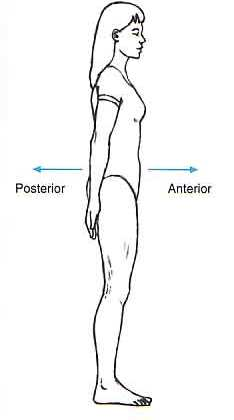
      • Anteriorly —1 frontal bone

      • Laterally — 2 temporal bones

      • Posteriorly — I occipital bone

      • Superiorly — 2 parietal bones

      • Inferiorly — I sphenoid and 1 ethmoid bone and parts of the frontal, temporal and occipital bones.

Fig. 1.1

  • Vertebral Cavity:

    • It contains “Spinal Cord.”

    • Boundaries:

      • Are made by bones of the vertebral column i.e. by vertebrae  and the intervertebral discs between the bodies of the vertebrae.

  1. Ventral Body Cavities:

  • They are present on the ventral side of the body.

  • They are much larger in size as compared to the dorsal body cavities.

  • They are further classified as followings,

    • Thoracic Cavity

    • Abdominal Cavity.

    • Pelvic Cavity.

  • Thoracic Cavity: 

    • It is formed by,

      • Anteriorly — the sternum and costal cartilages of the ribs

      • Laterally — 12 pairs of ribs and the intercostal muscles

      • Posteriorly — the thoracic vertebrae and the intervertebral discs between the bodies of the vertebrae.

      • Superiorly — the structures forming the root of the neck

      • Inferiorly — the diaphragm, a dome-shaped muscle.

    • Contents

      • The main organs and structures contained in the thoracic cavities are (Fig. 1.2):

        • the trachea, 

        • 2 bronchi, 

        • 2 lungs

        • the heart, 

        • aorta, 

        • superior and inferior vena cava,

        • numerous other blood vessels

        • the oesophagus

Fig. 1.2

  • The thoracic cavity is divided into three parts,

    • Pleural Cavity: One around each lungs, Contains fluid.

    • Pericardial Cavity: Around heart, Contains fluid.

    • Mediastinum: Central part extends from sternum to vertebral column and from first rib to diaphragm, contains all organs of thoracic cavity except lungs.

  • Abdominal Cavity:

    • This is the largest cavity in the body and is oval in shape (Fig 1.3).

    •  It is situated in the main part of the trunk and its boundaries are:

      • Superiorly — the diaphragm, which separates it from the thoracic cavity 

      • Anteriorly — the muscles forming the anterior abdominal wall 

      • Posteriorly —the lumbar vertebrae and muscles forming the posterior abdominal wall 

      • Laterally — the lower ribs and parts of the muscles of the abdominal wall 

      • Inferiorly — the pelvic cavity with which it is continuous.

Fig 1.3

The abdominal cavity is divided into the nine regions shown in Figure 1.4. 

Figure 1.4

  • Contents

    • the stomach, 

    • small intestine and most of the large intestine

    • the liver, gallbladder, bile ducts and pancreas.

    • the spleen

    • 2 kidneys and the upper part of the ureters

    • 2 adrenal (suprarenal) glands

    • numerous blood vessels

    • lymph vessels,

    • nerves

    • lymph nodes.

  • Pelvic Cavity:

    • The pelvic cavity is roughly funnel shaped and extends from the lower end of the abdominal cavity (Fig 1.4). 

    • The boundaries are:

      • Superiorly — it is continuous with the abdominal cavity 

      • Anteriorly — the pubic bones 

      • Posteriorly — the sacrum and coccyx 

      • Laterally — the innominate bones 

      • Inferiorly — the muscles of the pelvic floor.

Fig 1.4

  • The pelvic cavity contains the following structures: 

    • sigmoid colon, rectum and anus 

    • some loops of the small intestine 

    • urinary bladder, lower parts of the ureters and the urethra

    •  in the female, the organs of the reproductive system: the uterus, uterine tubes, ovaries and vagina.

    • in the male, some of the organs of the reproductive system: the prostate gland, seminal vesicles, spermatic cords, deferent ducts (vas deferens), ejaculatory ducts and the urethra.

Commonly Asked Questions:

  1. Enlist different body cavities along with their locations.

  2. Write in short about,

    1. Thoracic Cavity.

    2. Abdominal Cavity.

    3. Pelvic Cavity.

Labels:

1/09/2021

Basic Anatomical Terminologies.

  Introduction to the Human Body: Basic Anatomical Terminologies.

Introduction:

  • Human beings are the most complex of all the living organisms.

  • Human body contains many organs which work in an organized manner for an efficient working of the human body.

  • The study of human body is divided broadly in two divisions,

    • Anatomy

    • Physiology.

  • Human Anatomy is the branch of science that deals with the study of structures of different body parts.

  • Human Physiology is the branch of science that deals with the study of functions of different body parts.

Human Anatomy: Study of Structure.

Human Physiology: Study Of Functions.

Basic Anatomical Terminologies:

  • The universal terminologies are used to denote the body parts they are described as follows,

Body Positions:

  • Anatomical positions universally describe the regions and spatial relationships of the human body.

  • In anatomical position the body is “Upright” (The feet are placed on the floor flat and directed forward, the hands are at the sides with the palms facing forward as shown in the following figure.

  • Supine Position: 

    • Body lying flat on back, knees straight and hands at side as shown in the figure 1.1.

Figure 1.1: Supine Position

  • Prone Position:

    • Body lying flat on stomach, knees straight and hands at side as shown in the figure 1.2.

Figure 1.2: Prone Position

Directional Terminology used in Anatomy.

  • These terminologies are used to describe the position of one body part relative to another one.


Sr No.

Directional Term

Definition

Example

1

Superior

Towards the head or upper part of the body.

Pharynx is superior to larynx.

2

Inferior

Away from the head or lower part of the body.

Small intestine is inferior to stomach

3

Anterior

Nearer to or at the front of the body

Sternum is anterior to the heart.

4

Posterior

Nearer to or at the back of the body

Oesophagus is posterior to trachea.

5

Medial

Nearer to the midline of the body.

Ulna is medial to the radius.

6

Lateral

Nearer from the midline of the body.

The arms are lateral to the chest.

7

Intermediate

Between the two structures.

Transverse colon is intermediate to ascending colon and descending colon.

8

Proximal

situated nearer to the point of attachment.

Humerus is proximal to the radius.

9

Distal

situated away from the point of attachment.

Phalanges are distal to the carpels.

10

Superior

Toward or on the surface of the body.

Ribs are superficial to lungs.

11

Deep / Inferior

Away from the surface of the body.

Ribs are deep to the skin.


Planes.

  • Body planes are imaginary flat surfaces that pass through the body.

  • Sagittal, frontal and transverse planes are at right angle to one another.

  • The anatomical planes are as follows,

    • Sagittal plane.

    • Frontal plane.

    • Transverse plane.

    • Oblique plane.

  • Sagittal Plane:

    • It is a vertical plane that  divides the body into left and right sections.

  • Midsagittal (median) plane :

    • divides the body into equal halves at midline. 

  • Parasagittal plane :

    • Does not pass through midline, divides the body into unequal halves. 

  • Frontal (coronal) plane – 

    • divides the body or an organ into anterior and posterior sections.

  • Transverse (horizontal) plane:

    • divides the body into superior and inferior sections.  

  • Oblique Plane:

    • Pawsses through a body or an organ at an angle between transverse plane and sagittal plane or between a transverse plane and a frontal plane.

Sections:

  • A section is a cut made along the plane of the body.

  • There are three types of the sections,

  • Transverse Cut:

    • Section along the transverse plane, divides into anterior and posterior views.

  • Frontal Section:

    • Section along the frontal plane, divides into dorsal and ventral views.

  • Midsagittal Section:

    • Section along the midsagittal plane divides into right and left part.

Commonly asked questions.

  1. Explain different directional planes in the human body.

  2. Define with examples the following anatomical terminologies,

    1. Anterior.

    2. Posterior.

    3. Superior.

    4. Intermediate.

    5. Dorsal.

    6. Proximal.

    7. Distal.

    8. Medial.

    9. Lateral.

Labels: