Introduction:
The microbiological or microbial assay is a type of biological assay in which the relative potency of activity of a compound is determined by measuring the amount required for producing the predicted effect on a suitable test organism under standard conditions.
The microbiological assay of an antibiotic is based upon a comparison of the inhibition of growth of micro-organisms by measured concentrations of the antibiotics under examination with that produced by known concentrations of a standard preparation of the antibiotic having a known activity.
Two general methods are usually employed, the cylinder-plate (or cupplate) method and the turbidimetric (or tube assay) method.

Media Used for Antibiotic Assay:
Prepare the media required for the preparation of test organism inocula from the ingredients listed in Table 1.1
Dissolve the ingredients in sufficient water to produce 1000 ml and add sufficient 1 M sodium hydroxide or 1 M hydrochloric acid, as required so that after sterilization the pH is as given in Table 1.1
Table 1.1 Composition of media used for Anibiotic Assay (Quantity in gm per 1000ml)

Standard Preparation:
A Standard Preparation is an authentic sample of the appropriate antibiotic for which the potency has been precisely determined by reference to the appropriate international standard.
The Potency of the standard preparation may be expressed in International Units or in μg per mg of the pure antibiotic.
The Standard Preparations for India are certified by the laboratory of the Indian Pharmacopoeia Commission or by any other notified laboratory(ies) and are maintained and distributed by the agency(ies) notified for the purpose.
A Standard Preparation may be replaced by a working standard prepared by any laboratory which should be compared at definite intervals under varying conditions with the standard.
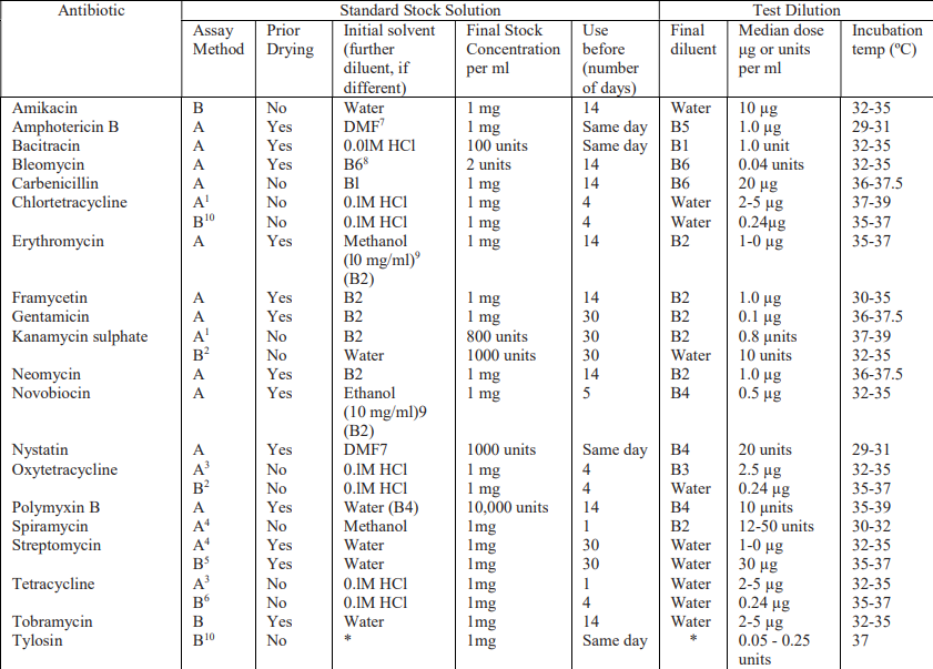
Dissolve a quantity of the Standard Preparation of a given antibiotic, accurately weighed and previously dried as indicated in Table 1.2, in the solvent specified in the table, and then dilute to the required concentration as indicated.
Store in a refrigerator and use within the period indicated.
On the day of assay, prepare from the stock solution five or more test dilutions, in the ratio 1:1.25 for Method A or smaller for Method B
Table 1.2 - Stock solutions and test dilutions of Standard Preparation.
Buffer Solutions.
Prepare by dissolving the following quantities given in Table 1.3 of dipotassium hydrogen phosphate and potassium dihydrogen phosphate in sufficient water to produce 1000 ml after sterilisation, adjusting the pH with 8 M phosphoric acid or 10 M potassium hydroxide.
Table 1.3 Composition of Buffer Solutions for Microbiological Assay of Antibiotics.

Preparation of the Sample Solution:
From the information available for the substance under examination (the “unknown”), assign to it an assumed potency per unit weight or volume, and on this assumption prepare on the day of the assay a stock solution and test dilution as specified for each antibiotic in Table 1.2 but with the same final diluent as used for the Standard Preparation.
Test Organisms:
The test organism for each antibiotic is listed in Table 1.4, together with its identification number in the American Type Culture Collection (ATCC).
Maintain a culture on slants of the medium and under the incubation conditions specified in Table 1.5, and transfer weekly to fresh slants.
Table 1.3 Test Organisms for Microbiological Assay of Antibiotics.

Preparation of inoculum:
For Method A:
After the suspension is prepared as given under Table 1.5, add different volumes of it to each of several different flasks containing 100 ml of the medium specified in Table 1.2.
Using these inocula, prepare inoculated plates as described for the specific antibiotic assay.
While conducting cylinder-plate assays, double-layer plates may be prepared by pouring a seed layer (inoculated with the desired micro-organism) over a solidified uninoculated base layer.
For each Petri dish, 21 ml of base layer and 4 ml of the seed layer may be generally suitable.
Fill each cylinder with the median concentration of the antibiotic (Table 1.2) and then incubate the plates.
After incubation, examine and measure the zones of inhibition.
The volume of suspension that produces the optimum zones of inhibition with respect to both clarity and diameter determines the inoculum to be used for the assay.
For Method B:
Proceed as described for Method A and, using the several inocula, carry out the procedure as described for the specific antibiotic assay running only the high and low concentrations of the standard response curve.
After incubation, read the absorbances of the appropriate tubes.
Determine which inoculum produces the best response between the low and high antibiotic concentrations and use this inoculum for the assay
Table 1.5 Preparation of Inoculum.

Methods of preparation of test organism suspension:
1. Maintain the test organism on slants of Medium A and transfer to a fresh slant once a week.
Incubate the slants at the temperature indicated above for 24 hours.
Using 3 ml of saline solution, wash the organism from the agar slant onto a large agar surface of Medium A such as a Roux bottle containing 250 ml of agar.
Incubate for 24 hours at the appropriate temperature.
Wash the growth from the nutrient surface using 50 ml of saline solution.
Store the test organism under refrigeration.
Determine the dilution factor which will give 25 per cent light transmission at about 530 nm.
Determine the amount of suspensions to be added to each 100 ml of agar of nutrient broth by use of test plates or test broth.
Store the suspension under refrigeration.
2. Proceed as described in Method 1 but incubate the Roux bottle for 5 days.
Centrifuge and decant the supernatant liquid.
Resuspend the sediment with 50 to 70 ml of saline solution and heat the suspension for 30 minutes at 70º.
Wash the spore suspension three times with 50 to 70 ml of saline solution.
Resuspend in 50 to 70 ml of saline solution and heat- shock again for 30 minutes.
Use test plates to determine the amount of the suspension required for 100 ml of agar.
Store the suspension under refrigeration.
3. Maintain the test organism on 10 ml agar slants of Medium G. Incubate at 32º to 35º for 24 hours.
Inoculate 100 ml of nutrient broth. Incubate for 16 to 18 hours at 37º and proceed as described in Method I.
4. Proceed as described in Method 1 but wash the growth from the nutrient surface using 50 ml of Medium 1 (prepared without agar) in place of saline solution.
Assay Methods:
Microbiological assays of Antibiotics are carried out using one of the following methods,
Method A: Cup-plate or Cylinder Plate Method.
Method B: Turbidimetric or Tube assay Method.
A. Cylinder-plate or Cup-plate method:
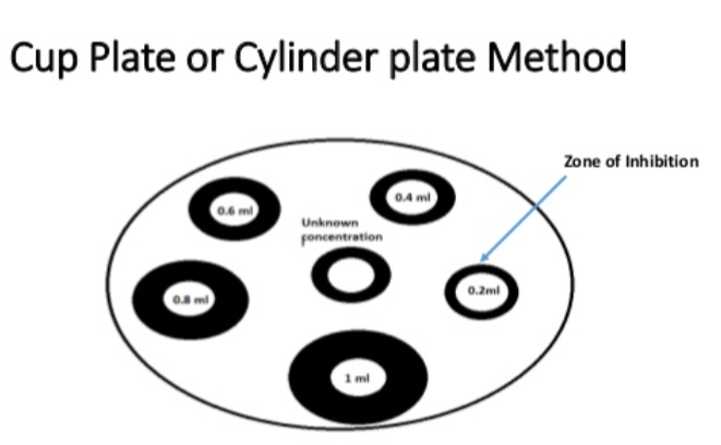
Principle: This method depends on the diffusion of antibiotic from a vertical cavity or cylinder through a solidified agar layer in a petri plate, which results in inhibition of growth of test microorganism in surrounding area of cavity or cylinder, the inhibition zone formed hence is corresponding to the potency of the antibiotic in use.
Inoculate a previously liquefied medium with the requisite quantity of suspension of the micro-organism, add the suspension to the medium at a temperature between 40º and 50º and immediately pour the inoculated medium into the petri dishes or large rectangular plates.
Spread test organism culture on it uniformly using spread plate technique.
Store the prepared dishes or plates in a manner so as to ensure that no significant growth or death of the test organism occurs before the dishes or plates are used and that the surface of the agar layer is dry at the time of use.
Solutions of known concentrations of the standard preparation and test antibiotic solution are prepared as per the guidelines given in table no. 1.2.
The volume of solution added to each cylinder or cavity must be uniform and sufficient almost to fill the holes when these are used.
When paper discs are used these should be sterilised by exposure of both sides under a sterilising lamp and then impregnated with the standard solutions or the test solutions and placed on the surface of the medium.
Leave the dishes or plates standing for 1 to 4 hours at room temperature or at 4º, as appropriate, as a period of preincubation diffusion to minimise the effects of variation in time between the application of the different solutions.
Incubate them for about 18 hours at the temperature indicated in Table 1.2. Accurately measure the diameters or areas of the circular inhibition zones and calculate the results.
Selection of the assay design should be based on the requirements stated in the individual monograph.
B. Turbidimetric or Tube assay method:
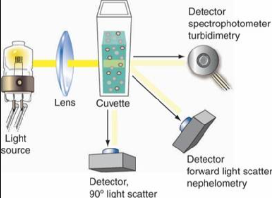
This method is not recommended for cloudy or turbid preparations.
Principal: Growth of microorganisms is calculated in the fluid medium supporting its faster growth in presence of an antibiotic.
The method has the advantage of a shorter incubation period for the growth of the test organism (usually 3 to 4 hours).
Prepare five different concentrations of the standard solution for preparing the standard curve by diluting the stock solution of the Standard Preparation of the antibiotic (Table 1.2) and increasing stepwise in the ratio 4:5.
Select the median concentration (Table 1.2) and dilute the solution of the substance being examined (unknown) to obtain approximately this concentration.
Place 1 ml of each concentration of the standard solution and of the sample solution in each of the tubes in duplicate.
To each tube add 9 ml of nutrient medium (Table 1.2) previously seeded with the appropriate test organism (Table 1.2).
At the same time prepare three control tubes, one containing the inoculated culture medium (culture control), another identical with it but treated immediately with 0.5 ml of dilute formaldehyde solution (blank) and a third containing uninoculated culture medium.
Place all the tubes, randomly distributed or in a randomized block arrangement, in an incubator or water-bath and maintain them at the specified temperature (Table 1.2) for 3 to 4 hours.
After incubation add 0.5 ml of dilute formaldehyde solution to each tube.
Measure the growth of the test organism by determining the absorbance at about 530 nm of each of the solutions in the tubes against the blank.