Cell Division: Mitosis
Cellular level of organization: Cell Cycle and Mitosis.
Introduction:
Cell is defined as a basic structural and functional unit of the body.
Cytology is a branch of science that deals with the study of cells.
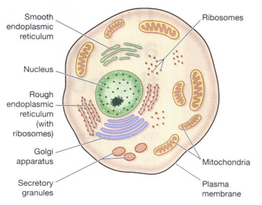
The animal cell is divided into two major parts as,
Plasma Membrane.
Subcellular organelles.
Nucleus.
Nucleolus
Mitochondria.
Golgi complex.
Ribosomes.
Endoplasmic reticulum.
Lysosomes.
Centrioles.
The cell cycle, or cell-division cycle, is the series of events that take place in a cell that cause it to divide into two daughter cells.
These events include the duplication of its DNA (DNA replication) and some of its organelles, and subsequently the partitioning of its cytoplasm and other components into two daughter cells in a process called cell division.
The events in the cell that include the duplication of its DNA (DNA replication) and some of its organelles, and subsequently the partitioning of its cytoplasm and other components into two daughter cells in a process called cell division.
Cell Division: The ability of the cell to produce daughter cells.
Normally, cells divide to replace dead or missing cells,
intestinal cells divide every 3 days, are broken down by digestion
blood cells last 3 months, are replaced by new cell division
nerve cells usually don't divide, last for life
Embryo: almost constant cell division. Every 30 minutes.
Sex cells are unique: in humans, males produce 500,000,000 /day. Females 1/month.
Cell Cycle:
It consists of following phases:
Interphase & Mitosis.
Interphase consists of:
G0 (Resting)
G1 (Gap 1)
G2 (Gap 2)
S (Synthesis)
M (Mitosis)
Cytokinesis (Separation)

Once a cell completes dividing (M), it enters interphase,
Interphase
Interphase is "normal" cell life -- it is busy making proteins and other polymers, housecleaning, and doing whatever its special functions require of it.
The stage following division (M), but preceding DNA synthesis (S) is called G1, or gap 1 phase. Actually, many cells sit in this phase for long periods of time, and some never leave it. Except for rapidly dividing cells, it makes more sense to think of this phase as Go, a point of exit from the cell cycle altogether.
At some point, the cell may be "triggered" to begin preparing for another round of cell division. This critical step commits the cell to DNA synthesis, during which every chromosome is replicated. At this point the cell leaves G1 (or Go). This fairly long process is called "S" phase, for synthesis.
Once S is completed, the cell continues to prepare for division, called the "G2", or gap 2 phase (gap between S and M).
Once mitosis begins, the cell is in the "M" phase. Mitosis consumes most cell energy for its duration, so much normal cell activity ceases.
The M phase terminates with cytokinesis, the physical separation of the two daughter cells.
Mitosis
Mitosis is a type of cell division that results in two daughter cells each having the same number and kind of chromosomes as the parent nucleus.
Stages of MITOSIS:
Interphase
Prophase
Metaphase
Anaphase
Telophase
Cytokinesis
Interphase:
cell is not dividing
cell is metabolically active
nucleolus visible, ribosomes being made
DNA duplicates (defines the S phase of interphase; before this is G1, after this is G2)
nucleus intact.

Prophase:
nucleolar material disperses
centrosomes move to opposite ends of cells.
mitotic spindle forms -- set of microtubules running from each pole to approximate the middle of the cell, a football-like structure.
chromatin condenses, chromosomes appear as pairs of identical sister chromatids, held together at centromere
kinetochores appear at centromere region (one for each chromatid)
chromosomes attach to kinetochore microtubules
nuclear membrane disappears
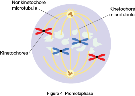
Metaphase:

microtubules from both poles manage to attach to kinetochores, pull chromosomes into a line at middle of cell = equatorial plate
anatomy of metaphase chromosome: two identical chromatids, essentially two separate chromosomes, but still attached at centromere.
Note: certain drugs (e.g. colchicine) block separation of chromosomes. All mitotic cells reach metaphase, stop = "metaphase arrest"
"Metaphase Plate '': Centrioles move to opposite poles while the chromosomes lie on the equatorial plane, centrioles form a full spindle the position is called as "Metaphase Plane".
Anaphase:
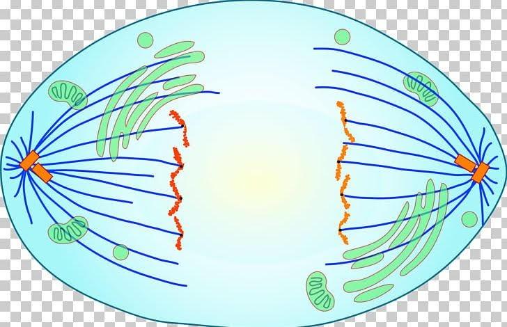
Spindle fibers start contracting.
Forms "V" Shape.
Sister chromatids start getting pulled towards opposite poles of the cell.
In anaphase actual division of chromosomes takes place.
Telophase:

chromosomes arrive at poles
chromosomes decondense, return to tangled mass of chromatin
spindle disappears
nuclear membrane reappears
nucleolus reappears, ribosome synthesis begins anew
Cytokinesis:
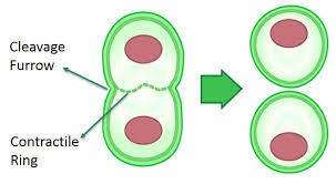
daughter cells are pinched apart.
Cleavage furrow grows and splits the cell into two daughter cells.
Commonly Asked Questions.
Define Cell DIvision.
Name different steps of cell cycle.
Name different steps of Mitosis.
Write a short note on Mitosis.
Labels: Human Anatomy and Physiology


<< Home